Q1. State the function of the cuticle.
Solution
The cuticle prevents the loss of water from the
surface of leaves.
Q2. What is the stomatal apparatus?
Solution
The stomatal aperture, guard cells and subsidiary
cells surrounding the guard cells are collectively called the stomatal
apparatus.
Q3. What are mesophyll cells?
Solution
Mesophyll cells are thin-walled chloroplast-containing
cells found in the ground tissue of leaves.
Q4. Differentiate between apical meristem and lateral
meristem.
Solution
Apical Meristem
Lateral Meristem
It
occurs at the tips of roots and shoots.
It
occurs in the mature regions of the roots and shoots.
They
produce primary tissues.
They
produce secondary tissues.
It
is responsible for the formation of young leaves and elongation of
roots and stems.
It
is responsible for producing a woody axis and for the thickness of the
plant.
Q5. Answer the following with respect to the arrangement of cells in a dicot stem.
Where are the medullary rays located?
What is the function of the hypodermis?
State the location of the pericycle.
Solution
Q6. Write the names of three simple tissues found in
plants.
Solution
Three simple
tissues found in plants are parenchyma, collenchyma and sclerenchyma.
Q7. Why do
the primary and secondary phloem get crushed during the activity of the
cambial ring?
Solution
During the activity of the cambial ring, the secondary xylem continues
to form and accumulate due to which the primary and secondary phloem get
crushed.
Q8. State
the type of vascular bundles seen in a monocotyledonous stem.
Solution
The type of vascular bundles seen in a monocotyledonous stem is of
conjoint and closed type.
Q9. Distinguish between the dorsiventral leaf and the isobilateral leaf.
Solution
Dorsiventral Leaf
Isobilateral Leaf
The number of stomata is more on the abaxial epidermis than the adaxial epidermis.
Almost equal number of stomata is present on the abaxial and adaxial surfaces.
Mesophyll is differentiated into spongy and palisade parenchyma.
Mesophyll layer is not differentiated into spongy and palisade parenchyma.
Vascular bundles are large and vary in size as per the size of veins.
Vascular bundles are similar in size, only the bundles near the mid vein are large.
Bulliform cells are absent.
Bulliform cells are present.
Q10. When is
the leaf surface in grasses exposed?
Solution
When the bulliform cells absorb water and become turgid, the leaf
surface in grasses is exposed.
Q11. Describe the sclerenchyma as a simple permanent
tissue.
Solution
Sclerenchyma is one
of the simple permanent tissues.
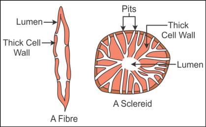 The cells are long,
narrow thick.
The cell walls are
lignified with numerous perforated pits.
The cells of sclerenchyma
are dead and without protoplast.
Sclerenchyma may be
in fibre form or in sclereid form.
The fibres are
thick walled, elongated and pointed cells occur in groups.
Sclereids are
spherical, oval or cylindrical.
Sclereids are
highly thickened dead cells with the narrow lumen,
Sclereids are found
in fruit walls of guava, nuts etc.
The sclerenchyma
provides the medical support to plants.
The cells are long,
narrow thick.
The cell walls are
lignified with numerous perforated pits.
The cells of sclerenchyma
are dead and without protoplast.
Sclerenchyma may be
in fibre form or in sclereid form.
The fibres are
thick walled, elongated and pointed cells occur in groups.
Sclereids are
spherical, oval or cylindrical.
Sclereids are
highly thickened dead cells with the narrow lumen,
Sclereids are found
in fruit walls of guava, nuts etc.
The sclerenchyma
provides the medical support to plants.
Q12. Give two examples of fruits which consist of
sclereids in their walls.
Solution
Pear and guava consist
of sclereids in their walls.
Q13. Define subsidiary cells.
Solution
The epidermal cells which lie near the guard cells
and have become specialised in their shape and size are called subsidiary
cells.
Q14. Name
the two tissues which are involved in secondary growth.
Solution
The two tissues involved in the secondary growth of plants are
vascular cambium and cork cambium.
Q15. Draw a diagram of the conjoint closed vascular bundle.
Solution

Q16. Describe the four elements of xylem.
Solution
The xylem is a
complex permanent tissue which conducts water and minerals from the roots to
the different parts of the plant.
It is also responsible
for the mechanical strength of the plant.
It is composed of
four elements - tracheids, xylem vessels, xylem fibres and xylem parenchyma.
1. Tracheids:
Tracheids are elongated tube-like structures.
They have thick cells with tapering ends and lignified cell walls.
The cells are dead and without protoplasm.
2. Vessels:
Vessels are cylinder-like structures made of many cells called vessel
members.
Cells possess a large cell cavity, and the cell walls are lignified.
They are interconnected to each other through the perforations in
their common walls.
3. Xylem Fibres:
They are with highly thickened walls and obliterated lumen.
They are either septate or aseptate.
4. Xylem Parenchyma:
The cells are living and thin walled.
Their cell walls are made of cellulose.
Xylem parenchyma is responsible for the radial conduction of water in
plants.
It also stores food in the form of starch or fat and substances such
as tannin.
Q17. Name
the following:
The layer which surrounds the vascular bundles
in a dicot leaf
The outermost cortical layer of a dicot stem
Solution
Q18. State the location of parenchymatous cells in primary stems and roots.
Solution
Parenchymatous cells are present in the cortex, pericycle, pith and medullary rays of the primary stem and roots.
Q19. Which meristem is the cylindrical meristem?
Solution
Secondary meristem
is the cylindrical meristem.
Q20. Give any one example in which the guard cells are dumb-bell shaped.
Solution
Grass
Q21. Distinguish between meristematic tissue and
permanent tissue.
Solution
Meristematic Tissue
Permanent Tissue
Cells
show great potential of cell division.
1. Cells have lost the capacity to divide.
The
cells are located at specific regions such as root tips, shoot tips
etc.
2. This type of tissues is present in
the entire plant body.
This
tissue is responsible for plant growth.
3. This tissue is involved in functions
such as conduction of water, minerals, food materials etc.
Q22. Describe the structural arrangement of collenchyma.
Solution
Collenchyma is a simple permanent tissue.
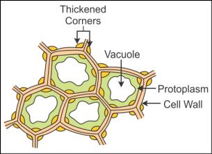 It is found either in the form of a homogeneous layer or in patches.
The cells are thickened at corners due to the deposition of cellulose, hemicelluloses and pectin.
Intercellular spaces are absent in collenchymas.
The cells are oval, spherical or polygonal.
They contain chloroplast and hence play role in assimilation of food.
It provides the mechanical support to the young growing parts of the plant.
It is found either in the form of a homogeneous layer or in patches.
The cells are thickened at corners due to the deposition of cellulose, hemicelluloses and pectin.
Intercellular spaces are absent in collenchymas.
The cells are oval, spherical or polygonal.
They contain chloroplast and hence play role in assimilation of food.
It provides the mechanical support to the young growing parts of the plant.
Q23. Describe with the help of a diagram the structure of a dicotyledonous root.
Solution
The outermost layer of the root is called the epidermis. Some of the epidermal cells protrude to give out root hair.
The cortex is made of several layers of thin-walled parenchymatous cells with intercellular spaces.
The innermost layer of the cortex is called the endodermis. It is made of large barrel-shaped cells without any intercellular spaces.
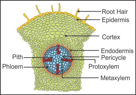 The tangential and radial walls of the endodermal cells have a deposition of water impermeable, waxy material called suberin in the form of casparian strips.
The pericycle, a layer of thick-walled parenchymatous cells, lies next to the endodermis.
The initiation of lateral roots and cambium occurs in the pericycle.
The pith is small and inconspicuous.
Between the xylem and the phloem is some parenchymatous cells known as conjunctive tissue.
Usually, there are two to four patches of xylem and phloem, and a cambium ring develops between them.
The tissue which lies on the inner side of the endodermis, i.e. pericycle, vascular bundles and pith, constitutes the stele.
The tangential and radial walls of the endodermal cells have a deposition of water impermeable, waxy material called suberin in the form of casparian strips.
The pericycle, a layer of thick-walled parenchymatous cells, lies next to the endodermis.
The initiation of lateral roots and cambium occurs in the pericycle.
The pith is small and inconspicuous.
Between the xylem and the phloem is some parenchymatous cells known as conjunctive tissue.
Usually, there are two to four patches of xylem and phloem, and a cambium ring develops between them.
The tissue which lies on the inner side of the endodermis, i.e. pericycle, vascular bundles and pith, constitutes the stele.
Q24. Enlist the elements of phloem.
Solution
The elements of
phloem are sieve tube elements, companion cells, phloem parenchyma and phloem
fibres.
Q25. What is
periderm?
Solution
The cells of the phellogen, phellem and phelloderm are collectively
called the periderm.
Q26. How many xylem bundles are present in a monocotyledonous root?
Solution
In a monocotyledonous root, there are more than six xylem bundles.
Q27. Describe the role of the cambial ring during the secondary growth in a dicotyledonous stem.
Solution
When the cambial ring becomes active, it starts to cut off new cells towards the inner as well as the outer side.
The cells which cut off towards the pith mature into the secondary xylem and the cells which cut off towards the periphery mature into the secondary phloem.
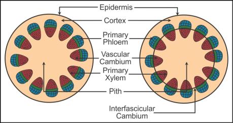
 Since the cambium is more active on the inner side, the amount of secondary xylem produced is more than secondary phloem.
The primary and secondary phloems get crushed due to the continued formation and accumulation of secondary xylem.
The primary xylem remains intact.
At some places, the cambium forms the narrow band of parenchyma which passes through the secondary xylem and phloem in radial directions.
These bands of parenchyma are called the secondary medullary rays.
Since the cambium is more active on the inner side, the amount of secondary xylem produced is more than secondary phloem.
The primary and secondary phloems get crushed due to the continued formation and accumulation of secondary xylem.
The primary xylem remains intact.
At some places, the cambium forms the narrow band of parenchyma which passes through the secondary xylem and phloem in radial directions.
These bands of parenchyma are called the secondary medullary rays.
Q28. State the functions of parenchyma.
Solution
Photosynthesis,
storage and secretion are the functions of parenchyma.
Q29. Explain the following terms:
Exarch
Endarch
Solution
Q30. Define tissue.
Solution
A tissue is a group
of cells which have a common origin and they usually perform a common
function.
Q31. Explain primary meristems and secondary meristems in
detail.
Solution
Primary
meristems:
There are two kinds
of primary meristems:
Apical meristems
Intercalary meristems
1. Apical meristems:
The meristems which occur at the tips of roots and shoots are called
apical meristems.
The root apical meristem is present at the tip of the root.
The shoot apical meristem is located at the distant most region of the
stem axis.
The apical meristem is responsible for the formation of young leaves
and elongation of stem and roots.
The shoot apical meristem gives rise to the axillary bud which may
form a new branch or a flower.
2. Intercalary meristems:
They occur between mature tissues.
They are found in grass. They help in the regeneration of parts
removed by the grazing animals.
Secondary Meristems:
They are also known as lateral meristems. They are cylindrical
meristems.
They occur in the mature regions of roots and shoots.
The secondary meristem is responsible for producing secondary tissues.
The fascicular vascular cambium, interfascicular cambium and cork cambium
are examples of the secondary meristem.
Q32. What are mature cells?
Solution
In primary and
secondary meristems, following division, the newly formed cells become
structurally and functionally specialised and lose the ability to divide.
Such cells are called mature cells.
Q33. State the function of companion cells.
Solution
The function of
companion cells is to maintain the pressure gradient in sieve tubes.
Q34. Name the following:
Thin-walled cells containing chloroplasts,
present in the ground tissue of leaves.
The vascular bundle common in stems and leaves.
Solution
Q35. Why are the cells of collenchyma much thickened at the corners?
Solution
Due to the deposition of cellulose, hemicelluloses and pectin, the cells of the collenchyma are much thickened at the corners.
Q36. Define
lenticels.
Solution
The phellogen cuts off the closely arranged parenchymatous cells on
the outer side instead of cork cells. These cells soon rupture the epidermis
to form a lens-shaped structure called lenticels.
Q37. What is
early bark and late bark?
Solution
The bark which forms in early season is called early bark, and the
bark which forms towards the end of the season is called late bark.
Q38. Explain the structure of stomata with a labelled diagram.
Solution
Structure of stomata:
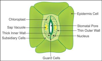 Stomata are present in leaf epidermis.
They regulate the process of transpiration and gaseous exchange.
They are enclosed by two bean-shaped guard cells.
The guard cells control the opening and closing of stomata.
Stomata are present in leaf epidermis.
They regulate the process of transpiration and gaseous exchange.
They are enclosed by two bean-shaped guard cells.
The guard cells control the opening and closing of stomata.
Stomata are present in leaf epidermis.
They regulate the process of transpiration and gaseous exchange.
They are enclosed by two bean-shaped guard cells.
The guard cells control the opening and closing of stomata.
Q39. Name the plants whose phloem fibres are of
commercial importance.
Solution
The phloem fibres
of jute, flax and hemp are of commercial importance.
Q40. What is
a conjunctive tissue?
Solution
A conjunctive tissue is a group of parenchymatous cells which lie
between the xylem and the phloem in a dicotyledonous root.
Comments
Post a Comment